2016 ~ Kedokteran dan Kesehatan

Friday, August 19, 2016

Hipothalamus Part IV

Neurotransmitters in the Hypothalamus

The hypothalamus has been referred to as a “pharmacological museum” (Goodman and Gilman,
‘55) by virtue of the plethora of neurotransmitters that it contains. Some of these neurotransmitters are found in the terminals of axons that originate from neurons outside the hypothalamus, but most are synthesized within the hypothalamus itself. The list of putative neurotransmitters includes the “classical” transmitters ACh, GABA, glutamate, serotonin, dopamine, and norepinephrine as well as literally dozens of peptides that have been identified in recent years.
While the overwhelming number of transmitters may create headaches for scientists and students, their presence offers hope for pharmacological intervention in the large number of different medical problems that are thought to involve the hypothalamus.


POINTS TO CONCENTRATE ON WHEN STUDYING:
1) Spatial relationships to surrounding brain structures. Don’t forget that the optic chiasm
   overlies the hypothalamus and therefore that visual disturbances (especially bitemporal
   hemianopsia, to be discussed in the vision lectures, can accompany pathology of the
   hypothalamus or hypophysis.

2) Know the location of each important nucleus or area with respect to the hypothalamic
   subdivisions (supraoptic, tuberal, etc.). This is, of course, helpful in diagnosis of lesions.

3) Functions: I consider all of the information in the handout with regard to functions as
   essential. However, you will not be tested on hormones since this material is covered in
   other courses. Read the section, “Functions of the Hypothalamus” very carefully.

4) Don’t worry about details in the “Neurotransmitters in the hypothalamus” section of the
   handout. Just read it for concepts.

Ginjal II (Proses Pembentukan Urin)

Proses Pembentukan Urin


  • Aliran darah ginjal (Renal Blood  Flow=RBF) diukur dengan
    •  - secara langsung         --> flowmeter
    • - secara tidak langsung--> metode klirens  (Clearance) Inulin, P.A.H dan Kreatinin
  • RPF = Jml zat yg diekskresi ginjal/mnt dibagi beda kdr zat dlm arteri & vena renalis
 Syarat zat untuk menghitung RBF (Renal Blood Flow) :
   - tidak dimetabolisme tubuh
   - disekresi (-) dan direabsorpsi (-)
   - tidak meracuni
   - tidak disimpan di ginjal
   - difiltrasi sempurna
   - mudah dianalisa

Ada 3 PROSES PEMBENTUKAN URIN

1. FILTRASI OLEH GLOMERULUS
2. REABSOPSI OLEH TUBULUS (terbesar di tubulus proksimal)
3. SEKRESI OLEH TUBULUS


PEMBENTUKAN URIN

  • Kecepatan ekskresi urin = laju filtrasi - laju reabsorpsi + laju sekresi
  • Zat difiltrasi bebas, mis : kreatinin
  • Zat difiltrasi bebas tetapi sebagian direabsorpsi , mis: elektrolit.
  • Zat difiltrasi bebas, ekskresi(-),semua direabsorpsi, mis : AA, glukosa
  • Zat difiltrasi bebas , Reabsorpsi (-), zat cepat dibersihkan dari darah dan diekskresi dalam jumlah besar di urin.

     FILTRASI GLOMERULUS

  • GFR = Kp ( (Pb. Pc) – a )

    Kp = konstante
    Pb = tekanan hidrostatik kapiler
    Pc = tekanan hidrostatik kapsula Bowmani
    a = tekanan koloid osmotik plasma
  • Kapiler glomeruli  kapiler lain 
    • Tekanan hidrostatik dipertahankan tetap
    • Permeabilitas tinggi (50x kapiler otot)
    • Luas permukaan glomerulus sangat luas
    • Filtrasi terjadi bila tekanan darah kapiler glomerulus > jumlah tekanan osmotik plasma dan tekanan kapsula Bowmani.
    • Jika tekanan darah aorta turun 40-50 mmHg urin tidak terbentuk.
    • BM < 70.000 dapat difiltrasi glomerulus
    • Klirens (Clearance) : jumlah plasma darah yang dibersihkan dari suatu zat oleh ginjal dalam satu  satuan waktu.

             Cx = Ux . V
                         Px


 SEKRESI DAN REABSORPSI TUBULUS

  • Reabsorpsi tubulus :
    • Tidak adanya zat tertentu dalam urin
    • Sedikitnya volume urin dibandingkan banyaknya filtrat glomerulus
  • Mekanisme Reabsorpsi dan sekresi tubulud : 
    • Pinositosis : protein, Asam amino
    • Transport pasif : air
    • Transport aktif  

       Reabsorpsi glukosa
  • Transport aktif
  • Normal kadar glukosa plasma : 80-120 mg%
  • Terjadi di permulaan tubulus proksimalis
  • Nilai ambang glukosa terhadap ginjal 300 mg%
  • Transport glukosa dalam tubulus tidak dipengaruhi insulin
  • Hampir semua reabsorpsi partikel larutan di tubulus proksimal, kecuali reabsorpsi Na pada semua tubulus renalis kecuali pada  pars ascendens ansa Henle. 

        Ekskresi air
  • Filtrasi glomerulus : 180 liter/hari
  • Urin yang terbentuk : 1 liter/hari
  • Urin pekat : air ditimbun melebihi larutan (garam).
  • Urin encer : air dilepaskan dari tubulus melebihi larutan garamnya
  • Reabsorpsi aktif di bagian permulaan tubulus proksimalis bersifat isotonis
  • Pars ascendes ansa Henle impermeabel terhadap air, Na aktif dipompa keluar dari filtrat larutan bersifat hipertonis
  • Filtrat di tubulus distalis bersifat hipotonis.
  • Osmolalitas dan volume filtrat di tubulus distali tergantung pada vasopresin menaikkan permeabilitas sel tubulus  distalis, duktus kolektivus terhadap air menyebabkan  urin sedikit dan pekat.

  Mekanisme “COUNTER-CURRENT”
  • Mekanisme pemekatan urin = perbedaan kenaikkan osmolalitas menyebabkan aktivitas ansa henle sebagai  “counter-current multiplier dan vasa rekta sebagai “counter-current exchanger” dan kemudian proses pasif tergantung pada difusi air dan Na 

   Faktor-faktor yang mempengaruhi  konsentrasi urin
  • Diuresis air menyebabkan kenaikkan tekanan osmotik efektif plasma memacu ekskresi vasopresin (ADH).
  • Intoksikasi air menyebabkan diuresis maksimal (aliran urin 16 ml/menit > minum >> lama plasma hipotonis > intoksikasiair
  • Ekskresi ureum menyebabkan pemekatan urin terhadap zat non urea lbh baik bila tdk ada ureum.
  • GFR: GFR menurun menyebabkan urin lebih pekat 

 Ekskresi Na dan Cl serta pengaturannya
  • Na difiltrasi dalam jumlah besar oleh glomerulus, direabsorpsi di seluruh tubulus renalis kecuali di pars ascenden ansa Henle. Normalnya 90-99% filtrat Na direabsorpsi kembali.
  • Ekskresi Na sangat dipengaruhi GFR, juga oleh mineralokortikoid, Angiotensin II, Oksitosin, N. renalis
  • Sebagian besar ion Na direabsorpsi bersama dengan Cl, sebagian lagi direabssorpsi dgn cara : 1 ion Na  ditukar  dg 1 ion K atau ion H 

       Ekskresi ion K
  • Sebagian besar ion K yg difiltrasi, direabsorpsi kembali di tubulus proksimal, diekskresi oleh tubulus distalis.
  • Ekskresi ion K menurun jika ;
    • Jumlah ion K dalam tubulus distalis menurun
    • Sekresi ion H naik

       Sekresi ion H
  • Ion H dibentuk dari disosiasi , dicurahkan ke dalam lumen tubulus untuk ditukarkan dg ion Na
  • Sel tubulus mengandung karbonik anhidrase yg mempercepat pemecahan H2CO3 > H+  + HCO3-
  • pH batas dimana dapat terjadi sekresi ion H secara transport aktif adalah 5,5 

   REFLEKS BERKEMIH
  • Refleks berkemih adalah refleks medula spnalis yang seluruhnya bersifat autonomik, tetapi dapat dihambat atau dirangsang oleh pusat dalam otak.
  • Pusat-pusat ini antara lain : pusat perangsang dan penghambat kuat dalam batang otak, terutama terletak di pons, dan beberapa pusat yang terletak di korteks serebral yang terutama bekerja sebagai penghambat tetapi dapat menjadi perangsang 
  • Refleks berkemih merupakan dasar penyebab terjadinya berkemih, tetapi pusat yang lebih tinggi normalnya memegang peranan sebagai pengendali sebagai berikut :
  • Pusat yang lebih tinggi menjaga secara parsial penghambatan refleks berkemih kecuali jika peristiwa berkemih dikehendaki.
  • Pusat yang lebih tinggi dapat mencegah berkemih, bahkan jika refleks berkemih muncul, dengan membuat kontsraksi tonik terus menerus pada sfingter eksternus kandung kemih samapi mendapatkan waktu yang baik untuk berkemih.
  • Jika tiba waktu berkemih, pusat kortikal dapat merangsang pusat berkemih sakral untuk membantu mencetuskan refleks berkemih dan dalam waktu bersamaan menghambat refleks sfingter eksternus kandung kemih sehingga peristiwa berkemih dapat terjadi.
  • Berkemih di bawah keinginan tercetus dengan cara sebagai berikut :
  • Seseorang secara sadar mengkonstraksikan otot-otot abdomennya, yang meningkatkan tekanan dalam kandung kemih dan mengakibatkan urin ekstra memasuki leher kandung kemih dan uretra posterior di bawah tekanan, sehingga meregangkan dindingnya. Hal ini menstimulasi reseptor regang, yang merangsang refleks berkemih dan menghambat sfingter eksternus uretra secara simultan. Biasanya seluruh urin akan keluar, terkadang lebih dari 5 – 10 mL urin tertinggal di kandung kemih. 


SOURCE : FK UMY

Obesity Part II

PATHOLOGI CONSEQUENCES OF OBESITY


Obesity results in morbidity and mortality largely because of its associati on with other diseases, including diabetes, cardiovascular disease, hypertension, sleep apnea, endometrial cancer, colon cancer, and gall bladder disease. Overall , in the United States, the excess mortality of obesity accounts for 300,000 deaths per year. It was estimated that the total spent for both weight reducti on and treatment of the consequences of obesity was $100 billion in the United States in 2001. This represents 5.5% to 7.0% of all medical expenses.

Diabetes

The increased risk for type 2 diabetes in individuals with obesity is considerable. In persons aged 20 to 44, obesity i s associated with a fourfold increase in the relative risk of diabetes. In a
study of a cohort of more than 50,000 U.S. male health professionals, the risk of diabetes correlated strongly with BMI. In men with a BMI of 35 or higher, the multivariate relative risk of diabetes was 42.1 compared with the risk in men with a BMI of l ess than 23. BMI appears to be the dominant risk factor for type 2 diabetes. Even men with average relative weight had a significant increase in risk when compared with men in lower weight groups. A similar increased risk exists for women. Among 43,581 women enrolled in the Nurses' Health Study, the relati ve risk for type 2 diabetes at the 90th percentile of BMI
was 11.2 . Weight was the single most important predictor of diabetes. After adjustment for BMI, lack of exercise and a poor diet (i .e., foods with a high glycemic index and high in trans fat) were also associated with increased risk of diabetes. Another study examined new diagnoses of diabetes in a population between 18 and 44 years of age and found an inverse correl ati on with age and BMI. Adul ts developing
diabetes before age 44 had an average BMI of 39, whereas adults developing diabetes at 45 or older had an average BMI of 33. Among all adults, the odds ratio for developing diabetes is 6.38 for those with a BMI greater than 40. The results of these and other studies lend support to the concept that the vast majority of cases of type 2 diabetes could be prevented by the adoption of therapies and lifestyle characteristics that
decrease obesity.

Although the precise mechanism by which obesity contributes to insulin resistance and type 2 diabetes has not yet been defined, it is likely related to the producti on of various factors derived from the adipocyte that act on fat, liver, or muscle to impair insulin action. Obesity is itself associated with
hyperinsulinemia, and insulin may induce insulin resistance
through down regulation of the insulin receptor. Potential candidate substances produced by fat that may cause insulin resistance include tumor necrosis factor and other cytokines,
such as interleukin -6, and resistin and adiponectin. Increased
levels of free fatty acids are also capable of inhibiting insulin action. It is intriguing that a recent report found that treatment with high-dose salicylate markedly improved insulin resistance, suggesting that obesity may induce an inflammatory state that contributes to insulin resistance.

Cardiovascular Disease

Obesity is an independent risk factor for cardiovascular disease, including coronary artery disease and congestive heart failure, in both men and women. Waist-to-hip ratio is the best predictor, and it i s noteworthy that increased waist -to-hi p ratio has an effect in women even at the relatively low BMI of 25.
Visceral obesity is associated with increased occurrence of hypertension and an atherogenic lipid profile, both of which contribute to the development of cardiovascular disease. In addition, in the obese state, there is a need for perfusion of a greater mass of tissue, resulting in an increase in cardiac work. Blood volume, stroke volume, and cardiac output are all increased and result in increased ventricular mass, which is
reversible with weight loss.

Pulmonary Disease

Abnormalities in pulmonary function may be seen in obese
patients. These range from quantitative abnormalities in pulmonary function tests that have no
established clinical significance to major dysfunction replete with symptoms and morbid consequences. The increased metabolic rate in obese subjects increases O2 consumption and CO2 production, and these changes result in increased minute ventilation. In subjects with marked obesity, the compliance of
the chest wall is reduced, the work of breathing i s increased, and the respiratory reserve volume and vital capacity are reduced; a resultant mismatch between ventilation and perfusion may result in hypoxemia. Severe obesity may cause hypoventilati on, defined by the development of CO 2 retention. The full designation of the obesity-hypoventilati on, or pickwicki an, syndrome includes somnolence, lethargy, and
respiratory acidosi s and typicallyal so includes sleep apnea. Such patients may have reduced ventilatory drive to hypoxia and hypercapni a, as well as obstructive or mechanical causes of hypoventilati on, and sleep studies may be necessary to distinguish among these.

Gallstones

Obesity is associated with enhanced biliary secretion of cholesterol . This results in supersaturation of bile and a higher incidence of gallstones —particularly cholesterol gallstones. Fasting, as opposed to more limited caloric restriction, increases the saturation of bile by reducing the phospholipid component, and cholecystitis induced by fasting is a well -recognized problem in obese individual's.

Cancer

Excess weight has been associated wi th increased rates of cancer. A recent study examining data for more than a million patients enrolled in the Cancer Prevention Study demonstrates convincingly that obese individuals are at increased risk for a number of cancers. The most dramatic increase in risk is seen for liver cancer. The relative risk of liver cancer was almost 2-fold hi gher in men with a BMI of 30.0 to 34.9 than in
normal -weight individual's, an d it was 4.5-fol d higher in men with a BMI greater than 35. In men with a BMI higher than 35, the risk of stomach cancer was increased 1.94 -fol d, that of kidney cancer was increased 1.7 -fol d, and that of esophageal cancer was increased 1.6-foldover the risk in normal -weight individual's. The effect of obesity on cancers of the gastrointestinal tract was not as great in women, but the increase in relative risk i n women was the same as that in men for kidney cancer. In women with a BMI greater than 35, the relative risk of cancer of the uterus was 2.8, of cancer of the
cervix was 3.8, and of breast cancer was 1.7.

Thursday, August 18, 2016

Hipothalamus Part III

Mammillary Region

       The mammillary part of the hypothalamus consists of the posterior hypothalamic nucleus and the
       prominent mammillary nuclei. The posterior nucleus is a large, ill-defined group of cells that
       may play a role in thermoregulation (see below). The mammillary nuclei are considered to be
       part of the hypothalamus on anatomical grounds, but, unlike the other hypothalamic nuclei, they
       do not appear to be closely related to autonomic and endocrine functions. Instead, the
       mammillary nuclei are believed to play a role in memory, and will be discussed during the limbic
       system lecture. For now, just associate the mammillary nuclei with memory.

       Summary of hypothalamic fiber tracts: There are four major fiber systems in the hypothalamus,
       all of which are bi-directional. The two largest and most clearly defined tracts carry fibers to and
       from the mammillary nuclei: the fornix (Figs. 6 & 9; also see Fig. 6 in Olfactory Pathways and
       Limbic System handout) carries fibers to these nuclei from the hippocampal formation (to be
       described in the limbic system lecture), and the mammillothalamic tract contains fibers running
       to and from the anterior nuclear group of the thalamus (Figs. 5 and 9). The stria terminalis (Fig.
       6) is an afferent pathway to the hypothalamus from the amygdala (also to be described in the
       limbic system lecture). The medial forebrain bundle (Figs. 6, 9) is an ill-defined bundle of
       unmyelinated and thinly myelinated axons which courses through the lateral hypothalamic area.
       This bundle contains a very large number of different ascending and descending components (50
       were distinguished in a recent paper). These include fibers ascending from the monoamine cell
       groups in the brainstem, descending fibers from the olfactory cortex that may play a role in
       appetite control, and descending fibers from other basal forebrain structures. It has been
       implicated in controlling many physiological functions like sleep, and behavioral traits like
       depression and pleasure.

       Functions of the Hypothalamus: Many of the functions of the hypothalamus are of a homeostatic
       nature (maintenance of constant body states). Several of these have already been described in
       conjunction with the description of the nuclei involved. Such functions require the integration of
       activity in many different body systems. A good example is temperature regulation (see Fig. 7
       but do not learn this figure). When body temperature increases, neurons in the anterior part of
       the hypothalamus turn on mechanisms for heat dissipation that include sweating and dilation of
       blood vessels in the skin. When body temperature decreases, neurons in the posterior part of the
       hypothalamus are responsible for heat production through shivering, vasoconstriction in the skin,
       and blockage of perspiration. Lesions in the anterior part can result in hyperthermia (increase in
       body temperature) and lesions in the caudal part can result in hypothermia when the
       environmental temperature is low.

       A recent, very significant finding is a direct projection from the hypothalamus to the
       preganglionic sympathetic neurons in the lateral horn (intermediolateral cell column) of the
       spinal cord and to the preganglionic parasympathetic neurons in the dorsal motor nucleus of the
       vagus. (Recall from Dr. Harting’s lecture that Horner’s syndrome can result from interruption
       of the pathway from the hypothalamus to the lateral horn as it passes thru the brain stem.) The
       cells of origin for these projections are located in many different parts of the hypothalamus. The
       hypothalamus has long been known as the “head ganglion” of the autonomic nervous system, but
       had been assumed to mediate its effects through multisynaptic pathways.

       In addition to autonomic and endocrine functions, the hypothalamus is also involved in the
       control of emotional expression. As described above, bilateral lesions including the
       ventromedial nucleus of the hypothalamus in animals have long been known to produce
       expressions of rage (see Fig. 8 but do not learn this figure). Stimulation of various other
       hypothalamic nuclei produces a variety of other emotions including pleasure and fear.


It is important to bear in mind, however, that many other areas of the brain, especially those
collectively termed the limbic system (following lecture), are involved in the control of emotions.

Another aspect of hypothalamic function that is interesting to consider is that it may be the site
where, in many cases, emotional factors influence body functions. It is well known that many
functions that are under autonomic or hormonal control are influenced to a large degree by
emotions. For example, emotional factors can influence or block menstruation, lactation, or
sexual function. Since the hypothalamus is concerned with control of emotions, and regulation
of both hormone release and the autonomic nervous system, it is thought to be involved in the
mediation of such effects.

Lanjut ke Part IV

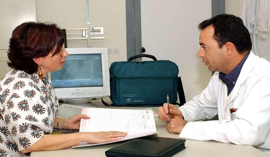
HISTORY_TAKING (ANAMNESIS)

ANAMNESIS

Tugas Utama Dokter :
·                     Membantu pasien mendapatkan derajat kesehatan yang optimal
·                     Memelihara kesehatan pasien agar selalu sehat
·                     Mendapatkan kesembuhan ketika pasien sakit
Prosedur Rutin Pemeriksaan Klinis
1. Anamnesis
2. Pemeriksaan Fisik
3. Pemeriksaan Penunjang (Laboratorium, Radiologi)
4. Diagnosis
5. Terapi
6. Edukasi dan konseling (follow up) serta prognosi
ANAMNESIS
·                Kegiatan awal dalam setiap tahapan pemeriksaan klinis
·                Anamnesis hubungan komunikasi antara dokter/tenaga kesehatan dengan pasien mengenai keadaan kesehatan pasien
·                Langkah yang sangat penting hampir > 80% dapat membantu menegakkan diagnosis
Physician-Centered
Physician's Agenda, Biomedical Focus dan Physician Gathers Data
Patient-Centered
Patient's Agenda, Symptom Focus dan Patient Tells Story
Your Role (as doctor)
·                     To gather the clinical history of the patient
·                     Done by being comfortable and profecient with patient interaction
·                     Collection of subjective and objective data
·                     Good questioning skill
Macam Anamnesis
1. Auto-anamnesis (dengan pasien sendiri)
 2. Hetero anamnesis/Allo-anamnesis (dengan orang yang dianggap mengerti tentang keadaan pasien)
Alloanamnesis
1.                   Penderita tidaksadar/koma
2.                   Penderita mengidap bisu/tuli atau afasia
3.                   Penderita bayi/anak
4.                   Penderita gangguan jiwa
Adapun alloanamnenis bisa menggunakan teknik kata-katayang mudah dipahami pasien.
Point-point anamnesis
1.                   Identitas pasien
2.                   Keluhan utama
3.                   Riwayat penyakit sekarang
4.                   Riwayat penyakit dahulu
5.                   Riwayat penyakit keluarga
6.                   Riwayat pribadi (personal sosial)
7.                   Review Anamnesis sistem
IDENTITAS
·                     Memberikan informasi : Siapakah penderita
·                     Masalah kesehatan yang mungkin muncul
·                     Mencari Faktor Resiko


IDENTITAS PASIEN
1.                  Nama :  Harus ditulis lengkap, manghindari kekeliruan, diusahakan nama sendiri
2.                  Umur : adanya penyakit dengan predisposisi timbul pada umur tertentu. Contoh : gondongan, campak (anak). Osteoporosis (wanita orang tua), degenratif (orang tua)
3.                  Jenis Kelamin : penyakit tertentu menyerang jenis kelamin tertentu. Contoh : osteoarthritis (wanita), gout (laki-laki). BPH (laki-laki), Ca Cervix (wanita).
4.                  Alamat : harus ditulis lengkap, hubungan dengan area epidomologi penyakit. Contoh : goiter (pembesaran kelenjar gondok) pada daerah pegunungan, malaria (daerah endemis missal Kulon Progo)
5.                  Agama dan Suku/RAS : Menghormati kebiasaan yang berkaitan dengan kegiatan keagamaan, budaya tertentu.
6.                  Pekerjaan : penyakit timbul karena akibat pekerjaan (occupational disease), atau sebagai pencetus penyakit. Contoh : Tuli ( tempat kerja bising  > 90 db), Pneumoconiasis ( pabrik tekstil, batu bara, abses). Alergi/asma (tempat berdebu)
7.                  Status Perkawinan : Belum cukup umur, menikah, janda/ duda
KELUHAN UTAMA
Keluhan yang dirasakan pasien/keluarga sangat mengganggu sehingga mendorong pasien/keluarga mencari pertolongan/nasihat medik. Misal : Demam, Nyeri kepala, muntah, Luka, diare, batuk, nyeri perut.
            Pada gangguan jiwa : sebab dibawa ke rumah sakit ( gaduh gelisah, mencoba bunuh diri)
RIWAYAT PENYAKIT SEKARANG
Riwayat perjalanan penyakit
·        Menggambarkan kronologis penyakit secara jelas dan lengkap
·        Dimulai dari akhir masa sehat dan awal masa sakit
Gejala Lengkap
1.      Lokasi : memnunjukan tempat keluhan pasien (missal di kepala, perut, dll)
2.      Waktu : menunjukan perjalanan penyakit
a.       Onset (kapan mulainya
b.      Durasi ( berapa lama)
c.       Seberapa sering (frekuensi)
3.      Kualitas : menunjukan karakter dari gejala. Misalnya : nyeri seperti terbakar, tajam, seperti ditusuk, menjalar, menekan. Batuk berdahak/kering.
4.      Keparahan : intensitas, kuantitas, atau meluasnya masalah. Misal : Jika nyeri dari skala VAS (0-10), jumlah lesi suatu kelainan kulit, dsb, batuk ngikil sampai muntah.
5.      Situasi dan kondisi saat terjadi (Faktor Pencetus) Meliputi faktor-faktor lingkungan, kegiatan personal, reaksi emosional atau situasi-kondisi yang lain yang berpengaruh ke keadaan sakit (mungkin meliputi alergi atau perilaku)
6.      Faktor-faktor yang memperberat atau meringankan gejala-gejala (remitting or exacerbating factors). Misalnya : dengan istirahat nyeri berkurang atau tidak ? Nyeri perut apabila diberi makanan tambah sakit ataukah berkurang
7.      Manifestasi gejala lain yang terkait. Gejala pada pasien tidak berhubungan dengan masalah kesehatan yang sekarang. Misal : Diare setelah terkena campak, batuk pilek disertai nyeri sendi.
SACRED SEVEN
a.       Lokasi
b.      Kualitas
c.       Kuantitas atau keparahan
d.      Waktu : Onset, durasi & frekuensi
e.       Situasi & kodisi saat terjadi
f.        Faktor-faktor yang memperberat atau meringankan gejala-gejala (remitting or exacerbating factors).
g.       Manifestasi gejala lain yang terkait

II. Riwayat Pengobatan
·        Pengobatan/Terapi yang sudah di dapat sebelumnya
·        Bagaimana hasilnya (ada perbaikan) ?
·        Etika ( Nama pribadi yang pernah menangani tidak perlu di tulis)

RIWAYAT PENYAKIT DAHULU
·        Riwayat penyakit baik fisik maupun psikiatrik yang pernah diderita sebelumnya. Misalnya riwayat trauma, penyakit serius lain,. Pembedahan, opname. Contoh : sering pusing, tidak bisa konsentrasi (riwayat CKB sebelumnya).
RIWAYAT KELUARGA

·        Penyakit dengan kecenderungan herediter, penyakit menular sering ditemukan dalam satu keluarga. Misal penyakit diabetes, darah tinggi, asma, alergi. Penyakit menular TBC, Lepra, dsb.
RIWAYAT PRIBADI
·        Riwayat Kehamilan
·        Riwayat Persalinan
·        Riwayat Imunisasi
·        Riwayat status perkawinan, hobby/kebiasaan  (alkohol, merokok) pola tidur, kondisi lingkungan rumah, tempat kerja
·        Riwayat alergi
Contoh Riwayat Personal
·        Prosmicuity (sering ganti-ganti partner) cenderung STD (penyakit menular seksual)
·        Kebiasaan merokok (ca paru)
·        Alkoholisme (sirosis hepatis/ kuning)
·        Kebiasaan tidur dengan bantalan tangan (hiperabduction syndrome)
REVIEW ANAMNESIS SITEM
·        Mencoba mengidentifikasi keluhan pada organ lain yang belum diungkapkan oleh pasien
Point-pointnya :
o   Keadaan umum : merasa lemah
o   Kepala/leher : nyeri kepala, leher kaku, mata (pandangan, kemerahan), telinga (berkurang pendengaran, berdenging, keluar cairan)
o   Sitem pernafasan : pilek (rinorhea), batuk (cough), sesak nafas (dyspnea), nyeri dada, batuk darah (hemoptoe)
o   Sistem Kardiovaskuler : dada berdebar (palpitasi), sesak nafas bila tiduran (ortopnea), malam hari terbangun karena sesak nafas (PND)
o   Sistem pencernaan : mual (nausea), nyeri perut (abdominal pain). Muntah (vomitus), muntah darah (hematemesis), berak hitam (melena), berak darah (hematocezia), diare, konstipasi, perut kembung (meteorismus)
o   Sistem Urogenital : nyeri kencing (disuria), anyang-anyangan (polaksuria), ngompol (enuresis), tidak dapat menahan kencing (inkontinensia), nyeri hebat di pinggang (kolic)
o   Sistem tulang dan otot : nyeri sendi (atralgia), nyeri otot (myalgia), deformitas, keterbatasan gerak
o   Sistem persarafan : separo anggota badan lemah (hemiparesis), lumpuh (hemiplegi), kesemuatan (paresthesia), kurang terasa (hypoesthesia), kebas (anesthesia) kehilangan kesadaran, gangguan daya ingat/memori, perhatian
Pencatatan Hasil
·        Hasil dari anamnesis dicatat di suatu blangko khusus yang sudah dirancang sebelumnya : Rekam Medis
·        Hendaknya dibuat selengkap mungkin (berguna dalam menyusun program rencana penanganan).
·        Data tersebut harus berupa pernyataan bukan hasil interpretasi.
·        Rekam medis merupakan dokumen rahasia, sehingga ada “wajib simpan rahasia kedokteran”
·        Merupakan kewajiban moril setiap tenaga kesehatan
·        Rekam Medis dilindungi hokum (undang-undang)